See The Human Brain Like Never Before
See The Human Brain Like Never Before
Authored by Makai Allbert via The Epoch Times (emphasis ours),
A decade ago, a small and unassuming brain sample arrived at Dr. Jeff Lichtman’s lab at Harvard University. Measuring less than a grain of rice, the 1 cubic millimeter of tissue contained 57,000 cells and 150 million synapses, each one a vital part of the brain’s intricate communication network.

Now, after a decade of collaboration with Google scientists, a monumental dataset—with 1,400 terabytes—has turned into the most detailed map of the human brain ever created.
“A terabyte is, for most people, gigantic, yet a fragment of a human brain—just a minscule, teeny-weeny little bit of human brain—is still thousands of terabytes,” Lichtman said in a National Institutes of Health report.
The detailed 3-D reconstruction reveals beautiful structures in the brain. Neurons forming dozens of connections, mirror-image neural pairs, and networks far more complex than expected, are just some of the groundbreaking discoveries.
“I remember this moment, going into the map and looking at one individual synapse from this woman’s brain, and then zooming out into these other millions of pixels,” said Viren Jain, a senior scientist at Google in Nature Magazine. “It felt sort of spiritual.”
The map, now part of an open-access dataset online, opens the door to new understandings of human cognition, psychiatric disorders, and the architecture of our minds.
“There is the saying that ‘A map of synaptic connections is necessary but insufficient to understand the brain.’ In its current form it is still missing a lot of important information, but it is a step in the right direction,” Daniel Berger, a scientist in the Lichtman lab, told The Epoch Times.
All images below are by Google Research and Lichtman Lab (Harvard University). Renderings by D. Berger (Harvard University)
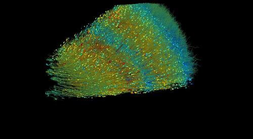
Excitatory neurons, color-coded by size, with red being the largest and blue the smallest. Cell cores range from 15 to 30 micrometers.
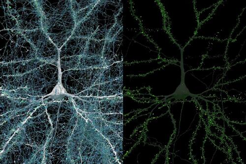
A single white neuron receives signals from more than 5,000 blue axons, with green synapses marking the points where the signals transfer.
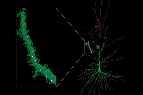
Neurons with long dendrites and dendrite spine. In very rare cases, a single axon (blue) made repeated synaptic connections (yellow) with a target neuron (green).
One unexpected discovery in the study was the presence of “axon whorls”—tangled loops of blue axons—which typically transmit signals away from nerve cells. These structures were rare in the sample and sometimes appeared to be resting on yellow cells. Their purpose remains unclear.
Read the rest here...
Related Posts
Wednesday Morning Minute
ories are trending at the moment and a look ahead at what the day may bring. Consider this your one-stop shop for news to kickstart your day. ]]>...
U.S. Chamber of Commerce congratulates Trump after 2021 rift
mmerce congratulated President-elect Donald Trump on Wednesday and pledged to work with his administration on deregulation and "pro-growth" policies. ...
Trump sees 'historic realignment' as GOP points to record Latino vote; gains across map
big for the GOP this week, giving the party arguably its best-ever numbers at the presidential level and hinting at a lasting political realignment. ...
Election Night Highlights: the Fun, the Joy, and the Meltdowns by the Media
as so many people came together to put President Donald Trump over the top to win the presidential race and become the 47th President-elect. ]]>...
WATCH: Kamala's Poor Response to Loss - Supporters' Faces Tell the Tale
ure we would be at this point on Tuesday night/Wednesday morning with the race called already, after all the talk of it possibly taking days. ]]>...


